
“That’s disgusting! Uhhh, don't do that!” his
voice was echoing in the farm office. Seventeen-year
old Ethan screamed when he heard me telling his dad that I had to get into
their piggery to gather some fresh pig placentas.
“Really?! What are you gonna do with it?” he
asked.
“I will have to ‘milk’ it so I could collect the placental umbilical cord blood in a tube and submit it to
the diagnostic laboratory,” I answered with a modulated voice hoping that this
young farmer with Asperger Syndrome could understand what I meant.
IT WAS A BRIGHT AND COOL early autumn morning at the Victoria-New South Wales border, I entered the farrowing (act of giving birth in pigs) room all geared up for the scheduled sample collection that Thursday. There was an increasing number of sows (mother pigs) in this piggery farm that could hardly get pregnant, the number of piglets born alive had been getting lower with higher rate of piglets born dead or mummified (we call it lan-os in our dialect).
Matthew—the farm owner,
who’s running the farm himself looked very frustrated at the office earlier
while he was telling me about his farm’s current reproductive performance. Their farm facility was relatively modern,
obviously well-updated to keep up with the demands of the pig farming
industry. Matt added that aside from
breeding issues, there had been an increasing number of dead pigs in the weaner
and grower sheds despite their regular improvements in husbandry, health and
nutrition.
Their consulting
veterinarian asked for a favour; he requested if our company could extend some diagnostic services so,
together, we could discover the real health issue of this piggery and eventually come up with a practical
and cost-effective solution for them. Having been
working with this pharmaceutical company that’s not just selling the product but offering some 'value' beyond the bottle was the main reason why I was, at that
moment, checking the farrowing crates of the piggery to see if there were fresh
afterbirths lying behind the newly-farrowed sows.
 |
| The placenta of a pig (diffused). Source: http://merrilysanimalsciencejournal.blogspot.com.au |
EVERY PIGLET BORN has
its own placenta, and because the average piglets born per sow at Matt’s Piggery were 9,
I could expect 9 placentas from each sow.
Disgusting as it may seem, the gelatinous placenta was actually the ultimate
reason why a piglet is born at full term— alive and healthy at the average of 114 days. Sadly, the placenta that gives life could potentially
get infected thus becoming an unwanted medium of disease transmission!
 |
| Human placenta, disc-shaped (DISCoidal). Source: Regeneration Center of Thailand |
Placental development in pigs starts 17 days after fertilization, and unlike the human baby’s placenta that attaches to the womb like a disc (discoidal), almost the entire surface (diffused) of the baby piglet’s placenta is attached to uterine wall of its mommy! And again, unlike in humans, the pig placental attachment is just a superficial connection of the lining of the foetal membrane that doesn’t create a deeper invasion into the inner wall of the sow’s womb (epitheliochorial).
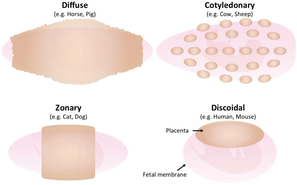 |
Placentation in animals, the shaded brownish area shows the attachment of the foetal membrane to the uterine wall.
Source: vle.du.ac.in |
Swine placenta is extra-gelatinous
compared to other animal species— it is a sac containing fluids and blood vessels
extending around the piglet to protect and nourish it. These tiny ducts and vessels gradually
converge together to become a bigger, highly functional sow-to-foetal piglet 'link' known as the umbilical cord that's approximately 12 centimetres (4.7 inches) in length! Since the lungs and intestines of a
developing piglet are not functional, the placenta, through meticulous blood filtration,
supplies oxygen and nutrients from the sow to the piglet via the umbilical cord.
 |
| Classification of placentas based on histological assessment of the maternal-chorion interface. Source: http://www.nature.com/ni/journal |
MY HEART LEAPT when,
finally, I spotted a sow lying and continuously grunting as if calling for her eight
groggy, pinkish and damp ‘babies’ scattered around the heated and well-lit farrowing
crate. While I was having fun observing
these cute little piglets as they obviously blinked, innocently root around and tried
to find their way up to their mum’s udder, I noticed some streaks of fresh, maroon
mucous staining the sow’s ham, tail and hock.
There was also a pile of placenta behind her. That’s what I exactly need, I almost
screamed at myself!
I asked for assistance
from the lovely farrowing room attendant.
Luckily she was happy to hold the red-topped blood tube for me as I
individually picked-up the slimy, expelled-inside-out afterbirths— I would
manually tear each one of them to expose and grab the umbilical cord stump... We were both double-gloved and wearing facial
mask and goggles to protect ourselves from the potentially contaminated splatters
but I could sense her revulsion to what I was doing especially when I uncovered
two mummified pig foetuses hidden under the pile of afterbirths!
I skillfully held each placenta, cut the clotted end of the navel cord off with a pair of scissors and desperately squeezed the blood out from the placental vessels through to the severed end of the cord that was comfortably resting inside our sampling tube. The process had to be repeated until I could come up with one substantial diagnostic sample— a collection of placental umbilical cord blood from 3-4 placentas sourced from one donor sow.
I skillfully held each placenta, cut the clotted end of the navel cord off with a pair of scissors and desperately squeezed the blood out from the placental vessels through to the severed end of the cord that was comfortably resting inside our sampling tube. The process had to be repeated until I could come up with one substantial diagnostic sample— a collection of placental umbilical cord blood from 3-4 placentas sourced from one donor sow.
 |
| A simple illustration of Placental Umbilical Cord Blood sampling. Illustrated by: Ruel P. Pagoto, DVM |
THE PLACENTAL UMBILICAL CORD
SERUM (PUCS) samples from a farm are the best diagnostic tools to establish the occurrence
of in-utero transmission of Porcine Circovirus 2 (PCV2). In addition to what has been commonly
observed in weaner and early grower pigs (emaciated, hairy, yellowish pigs with
varying sizes of red patches on their skin) around the globe, the infective
level of PCV2 in gilts (a young female swine), sows and boars could directly affect their reproductive
performance. This was the reason why we
decided to collect PUCS from Matthew’s Piggery; I submitted the precious samples
we collected to the diagnostic lab so they could check for the presence of viral
DNA for us.
Don’t worry; PCV2 virus doesn’t cause disease in humans so we’re safe.
In pigs, it can be transmitted via direct contact (nose-to-nose, mouth-to-nose), by contaminated needles, and through the boar’s semen! Unlike influenza virus, PCV2 virus is sort of ‘naked’ or non-enveloped, and its DNA contained in a naked capsid is single-stranded; but as it gets into the sow’s womb during breeding (via semen), this virus would acquire some necessary proteins from its host transforming itself into a double-stranded DNA virus— which makes it highly replicative! PCV2 will then divide and invade the sow’s blood stream (viraemia) thus having the potential of infecting the developing embryo at all stages of the reproductive cycle!
In pigs, it can be transmitted via direct contact (nose-to-nose, mouth-to-nose), by contaminated needles, and through the boar’s semen! Unlike influenza virus, PCV2 virus is sort of ‘naked’ or non-enveloped, and its DNA contained in a naked capsid is single-stranded; but as it gets into the sow’s womb during breeding (via semen), this virus would acquire some necessary proteins from its host transforming itself into a double-stranded DNA virus— which makes it highly replicative! PCV2 will then divide and invade the sow’s blood stream (viraemia) thus having the potential of infecting the developing embryo at all stages of the reproductive cycle!
 |
| The Porcine Circovirus2 before and after infecting a pig. Illustrated by: Ruel P. Pagoto, DVM |
If the embryo gets
infected on the early stage of development, embryonic resorption could occur. When PCV2 infection happens after foetal
bone development has commenced (35 days after fertilisation), it could
result into a mummified piglet! Luckily,
the piglet’s immune system (thymus) would start to develop but NOT fully functional
at 70 days of gestation (pregnancy), so when the foetal pig gets infected on or
beyond this period— the immune system would try to combat the infection, it may
survive, otherwise, a weak, sick or dead piglet is born.
 |
| PCV2-related reproductive failure. Source: Boehringer Ingelheim CircoFLEX brochure. |
Interestingly, as with
other diseases’ dynamics and severity, not all sows get infected with PCV2 at
the same time. Sows that get infected would
develop protective antibodies against the virus that are passed on to the newborn
piglets via colostrum; piglets of these infected sows are therefore protected but
could potentially shed the virus to the piglets born out of an uninfected sows that,
of course, haven’t developed specific protection! This could be the reason why increasing
number of weaners and young growers at Matt’s Piggery would still get sick and die
despite the updated health and nutrition program they’ve been implementing in
the farm.
 |
| PCV2 infection and immunity. Illustrated by Ruel P. Pagoto, DVM |
The lab test result was released 4 days after we submitted the samples. It's positive, trans-placental transmission was happening at the piggery farm!
We went back to the farm to give our recommended health program and Ethan was attentively listening to what we had to say during our meeting with his dad. It was clear to him that we were there to look after the health of their pigs. A ‘blanket’ PCV2 vaccination in boars and sows (regularly boosted twice a year) was implemented. All the incoming ‘maiden’ gilts had to be vaccinated 2 weeks before their entry to the breeding herd and the piglets, as it had been successfully implemented— PCV2 vaccination at 14 days or older.
WHEN WE TESTED the same herd eight months later, there were no DNA copies detected from the PUCS samples submitted. The farm’s performance had improved and the same health program was implemented to their other two pig breeding sites.
.
-----------------------------------------------------
References:
http://www.nda.agric.za/docs/Infopaks/Careofsowpiglets.pdf
http://www.thepigsite.com/pighealth/article/220/parturition-farrowing/
http://placentation.ucsd.edu/pig.html
Pig Diseases 9th edition by DJ Taylor
Swine Diseases 10th edition edited by Zimmermann et al.
Various publications by Boehringer Ingelheim


